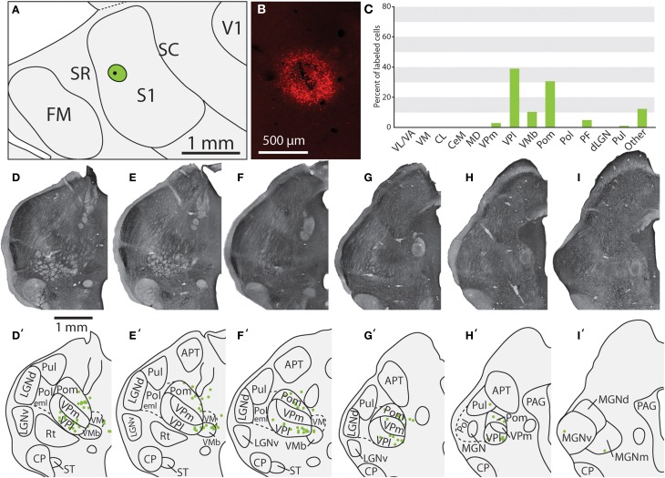
Figure 6.
Projections to the S1 body representation from in case 13-114. (A) Reconstruction of the flattened cortical hemisphere, showing the core of the injection site of FR (black dot) surrounded by the halo (green circle) in the body representation of S1. (B) Fluorescent image of the FR injection site and surrounding halo. (C) Percent of labeled neurons originating from various thalamic nuclei (D–I) Rostral to caudal progression of CO stained tissue; corresponding thalamic borders and labeled neurons are shown below (D'–I'). In this case, the majority of label was found within VPl, VMb, and Pom. All conventions as in previous figures.

