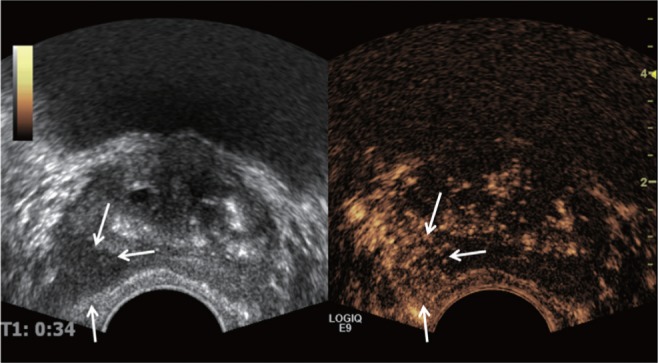
Fig. 2.

Contrast enhanced transrectal ultrasound (TRUS) findings of prostate cancer in a 62-year-old man. Contrast enhanced TRUS image shows increase vascularity and contrast agent signals from left peripheral zone suggesting increased vascularity (arrows). Note that the focal lesion shows low echogenicity in gray-scale TRUS, which is one of common findings of prostate cancer. This lesion was confirmed as prostate cancer after TRUS guided targeted biopsy.
