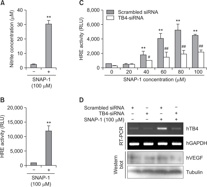
Fig. 2.
VEGF expression is upregulated by SNAP-1, NO donor. (A) HeLa cells were treated with 100 μM SNAP-1. NO production was detected as nitrite accumulated in culture supernatant by using Griess reagents. (B) HeLa cells were transfected with pGL2 plasmid of hypoxia response element (HRE)-luciferase (Luc) and treated with 100 μM SNAP-1. Luc activity was measured with luminometer using Luc substrate. Data in bar graph represent mean ± SED. **p<0.01, statistical significance vs. SNAP-1-untreated group (A and B). (C-D) HeLa cells were co-transfected with Tβ4-siRNA and pGL2-HRE-Luc plasmid. Then, cells were treated with various concentrations of SNAP-1. Luc activity was measured with luminometer using Luc substrate. Data in bar graph represent mean ± SED. Luc activity was measured with luminometer using Luc substrate. Data in bar graph represent mean ± SED. **p<0.01, statistical significance vs. SNAP-1-untreated group. #p<0.05; ##p<0.01, statistical significance vs. scrambled siRNA-treated group at each concentration of SNAP-1 (C). RNA was purified with TRIZOL reagent as described in materials and methods. Tβ4 transcript level was measured by RT-PCR and VEGF levels were detected by western blot analysis (D).

