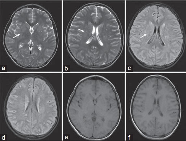
Figure 1.

16-year-old boy presented with altered sensorium of 1 day duration diagnosed with eosinophilic meningitis caused by A. cantonensis. MR images, (a and b) axial T2 (c and d) axial FLAIR images, at the basal ganglia level demonstrate subtle scattered hyperintensities in the periventricular regions (white arrows). (e and f) Axial T1-weighted images do not show these features.
