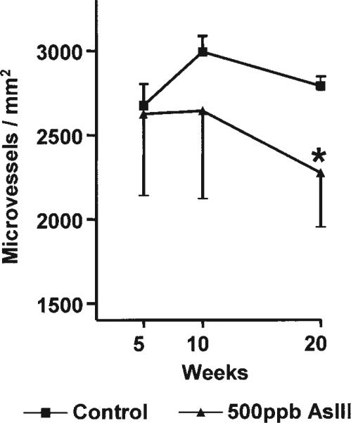
Fig. 7.
Chronic AsIII exposure reduced cardiac micro-vessel density. Middle ventricular sections of the hearts collected in Fig. 5 were formalin fixed, paraffin embedded, and cross-sectioned. The tissue was stained with Masson's trichrome and imaged at ×400. Microvessels were automatically detected using a digital imaging subroutine and vessels with cross-sectional diameters of <12 μm were enumerated and sums were normalized to tissue area in the same image. Each point represents the mean ± SD microvessel density from the hearts of five mice. Significant differences from time-matched control are designated by *p < 0.05.

