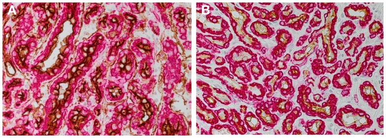
Figure 1.

DAB staining of proliferating infantile hemangioma showing the abundance of microvessels with an inner endothelium expressing the endothelial marker, CD34 [(A) brown], and an outer pericyte layer expressing smooth muscle actin [(A,B) red]. The inner endothelial layer also expresses GLUT-1 [(B) brown], the immunohistochemical marker for infantile hemangioma. Cell nuclei are counterstained with hematoxylin [(A,B) blue]. Original magnification 400×.
