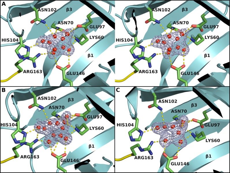
Figure 3.

The ligand binding site. The binding site is located at one end of the β-barrel as identified from binding of a UNL (red spheres). (A) Stereo view of the UNL and its interaction with the protein in BACOVA_00364 (pdb code 4gzv). Omit map is contoured at 1.25 σ level above the mean density. The residues interacting with the UNL are shown in stick representation. Arg163 that interacts with the UNL comes from an adjacent protomer in the biological tetramer. (B) and (C) are the corresponding UNL binding sites for BACUNI_03039 (pdb code 4iab) and BACEGG_00036 (pdb code 4i95), respectively, where the same or similar UNL is bound.
