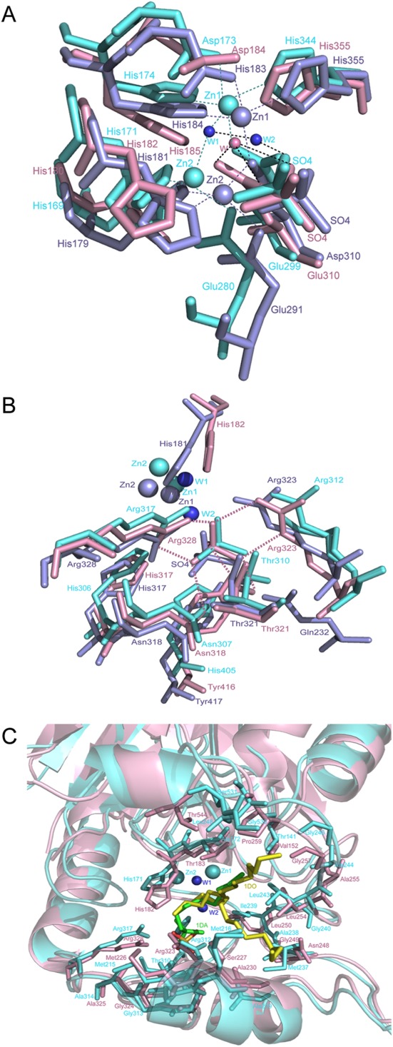
Figure 4.

Active site of YjcS. (A) Superimposition of the zinc binding site. YjcS is in light pink, SdsA1 is in aquamarine, and Pisa1 is in light blue. Two water molecules from SdsA1 are shown as small spheres in tv_blue and the water from YjcS is in light pink. Hydrogen bonds are indicated by dotted lines in black and the zinc coordination bonds are shown as dotted lines and coloured in aquamarine or light blue. (B) Superimposition of the sulfate group binding site. Hydrogen bonds between the sulfate group and residues in YjcS are marked as light pink dotted lines. (C) Superimposition of the YjcS structure with SdsA1 containing 1DA (green) and 1DO (yellow). The hydrophobic residues located in the substrate-binding site are shown as sticks.
