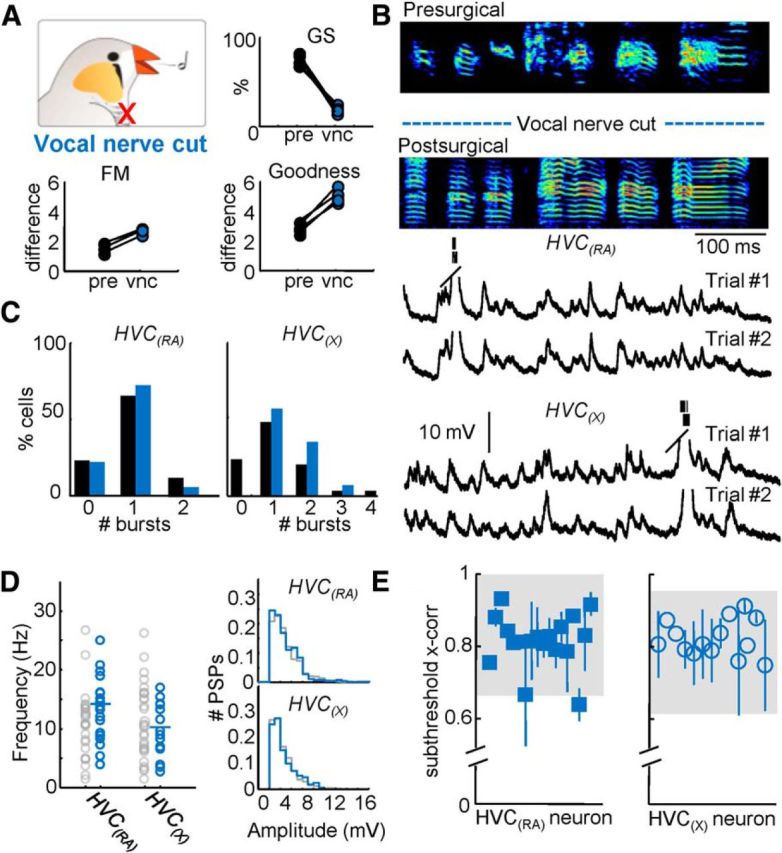
Figure 5.

Singing behavior and HVC projection neuron activity following vocal nerve transection. A, Top, Global similarity of four birds compared before surgery and after vocal nerve cut (vnc). Bottom, Difference of FM and goodness of pitch were significantly altered (p < 0.05) after vocal nerve cut. B, An example motif produced by the same bird before and after the vocal nerve was cut. Example intracellular recordings of an HVC(RA) and an HVC(X) neuron during two repetitions of the postsurgical motifs shown above. The postsurgical motif shown in B was produced while Trial #1 of the HVC(RA) neuron was recorded. C, Number of bursts per motif for both HVC(RA) and HVC(X) neurons. D, The mean frequency and amplitude distribution of identified excitatory synaptic inputs for HVC(RA) and HVC(X) neurons after vocal nerve cut. E, Subthreshold correlation coefficients were calculated for all cells recorded after vocal nerve cut (shaded region = 95% confidence interval).
