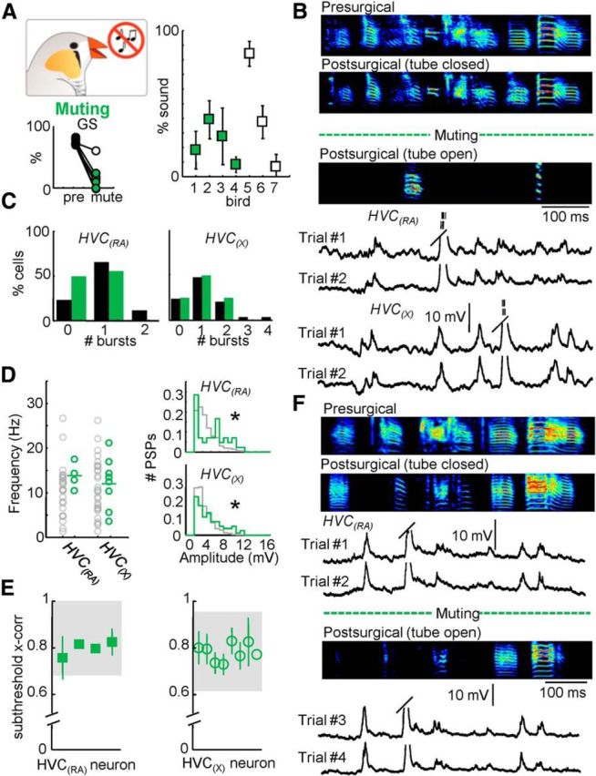
Figure 6.

Behavioral and electrophysiological effects of reversible muting. A, Percentage of presurgical song remaining after muting (n = 7 birds; right). Global similarity (GS) during normal and postsurgical (tube open) song production. B, Three motif examples from one bird highlighting the reversible muting approach: Presurgical (top); Postsurgical, tube closed (96% song remaining, middle); and Postsurgical, tube open (18% song remaining, bottom). Below the sonograms, example intracellular traces of an HVC(RA) and an HVC(X) neuron recorded during two repetitions of the postsurgical (tube open) motif are displayed. Trial #1 of the HVC(RA) neuron corresponds to the postsurgical (tube open) motif shown above. C, Number of bursts per motif for both HVC(RA) and HVC(X) neurons. D, The mean frequency and amplitude distribution of identified excitatory synaptic inputs for HVC(RA) and HVC(X) neurons after muting. E, Subthreshold correlation coefficients for all cells recorded after muting (shaded region = 95% confidence interval). F, Electrophysiological recording during reversible muting. Displayed are the presurgical song and two variants of the postsurgical song (tube open and tube closed). Four intracellular recordings from an HVC(RA) neuron are provided. Two of these examples are from the tube closed condition (Trials #1 and #2). After reversible muting (opening the tube), the bird sang the song displayed below the dotted line. Two examples of song-related membrane potential in the tube open condition are given below for that neuron (Trials #3 and #4).
