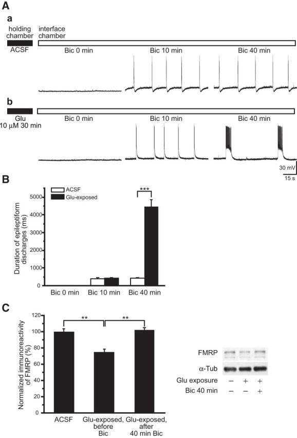
Figure 2.

Distinct prolonged epileptiform discharges were elicited in glutamate-exposed slices. Slices were maintained in a slice-holding chamber (filled horizontal bar) for glutamate exposure, and then transferred to an interface chamber for continuous bicuculline perfusion (empty horizontal bar). A, Representative intracellular recordings obtained at 0, 10, and 40 min following the addition of 50 μm Bic to the perfusion solution. Aa, In control slices, bicuculline elicited stable, short epileptiform discharges (Bic 10 min), and the short epileptiform discharges remained stable with continuous bicuculline perfusion (Bic 40 min). Ab, In slices exposed to glutamate (Glu 10 μm 30 min), short epileptiform discharges elicited by bicuculline (Bic 10 min) were followed by an abrupt appearance of prolonged epileptiform discharges that gradually dominated population responses (Bic 40 min). B, Average durations of epileptiform discharges following continuous bicuculline perfusion in control (ACSF, empty columns) and glutamate-exposed slices (filled columns). The average durations of initial epileptiform discharges at Bic 10 min in control and in glutamate-exposed slices were not significantly different. In contrast, at Bic 40 min the average duration of the epileptiform discharges in glutamate-exposed slices was significantly longer than that of control slices due to the occurrence of prolonged epileptiform discharges. C, FMRP levels in slices in ACSF, following glutamate exposure (Glu-exposed, before Bic), and after 40 min bicuculline in the recording chamber (Glu-exposed, after 40 min Bic). FMRP in ACSF was 100 ± 3.5%, n = 6; in Glu-exposed, before Bic: 75 ± 3.6%, n = 6; and in Glu-exposed, after 40 min Bic: 102 ± 2.5%; n = 6; one-way ANOVA; **p < 0.01).
