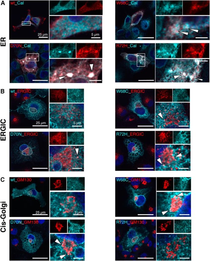
Figure 8.

Subcellular trafficking routes of α1 loop D/β2–3 mutants. A–C, GlyR α1 variants W68C, D70N, and R72H were analyzed for their localization in cellular compartments ER, ERGIC, and cis-Golgi. To exclude effects from overexpression, α1 variants were subcloned into a low-expression vector under the control of the ubiquitin promoter. A, Merged images showing costaining of GlyR α1 (red) with the ER protein calnexin (cyan, rabbit anti-calnexin, 1:500). Right, Magnification of the white rectangle with white arrowheads pointing to accumulated GlyRα1. B, Colocalization of α1 (cyan) with the ERGIC protein 53 (red, mouse anti-ERGIC53, 1:500); the ERGIC compartment is marked with a white dotted line and shown in higher magnification at top right. C, Costaining with the cis-Golgi marker GM130 (red, mouse anti-GM130, 1:500). Color codes are indicated in labels within the left images. The nuclei are stained with DAPI (blue). White arrows point to colocalization of GlyR α1 variants with compartmental marker proteins. Scale bars: 25 μm in left images, 5 μm for ERGIC, and 3 μm for cis-Golgi in enlarged compartment images.
