
Figure 3.
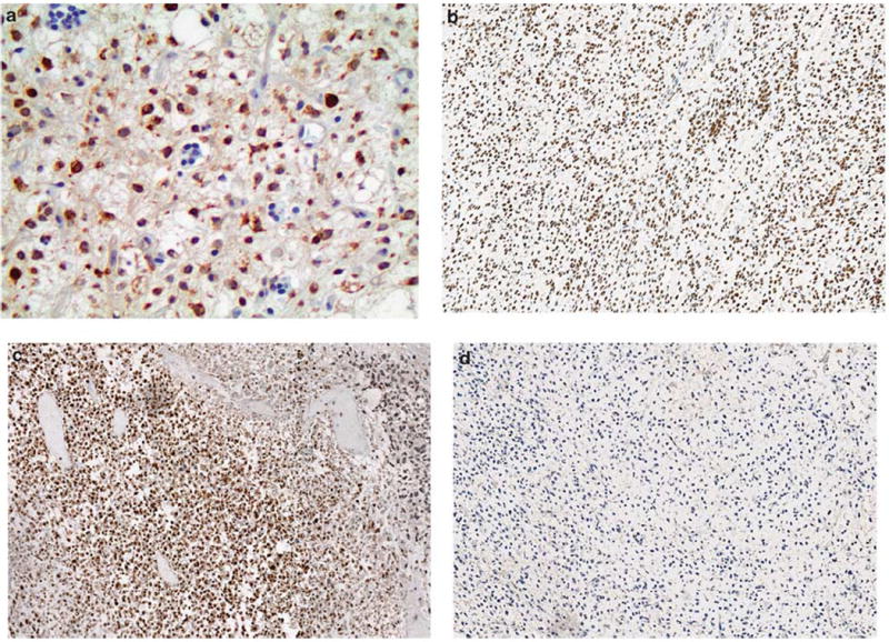
Cancer-testis antigen immunoreactivity in myxoid and round cell liposarcomas. (a) MAGEA1 (× 400), (b) PRAME (× 200), and (c) SSX2 (× 200), were expressed in varying proportions and demonstrate a predominantly nuclear staining distribution. (d) ACRBP (× 200) staining was negative in all samples.