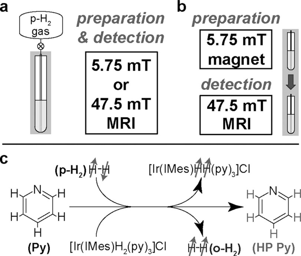
Figure 1.
Schematic diagram of the experimental setup for 1H NMR/MRI at low magnetic fields: a) In situ detection for which preparation (chemical exchange/polarization transfer) and detection were both performed in the same magnetic field (5.75 or 47.5 mT). b) Ex situ detection for which preparation (chemical exchange/polarization transfer) was performed at 5.75 mT and was followed by rapid transfer (≈ 8 s) to a 47.5 mT field for detection. Note that the ex situ approach primarily detects only one HP product (pyridine), whereas in situ detection allows “capturing” the HP product (pyridine) and two HP intermediates/byproducts (Ir–dihydride and orthohydrogen). No additional magnetic field (except that provided by Earth) was used during transfer of the sample from the 5.75 to 47.5 mT magnet for ex situ experiments. c) Molecular diagram of the exchange process between gaseous parahydrogen, the Ir complex, and pyridine to lead to the polarization transfer from parahydrogen (p-H2) to pyridine (Py), Ir–dihydride, and orthohydrogen (o-H2).

