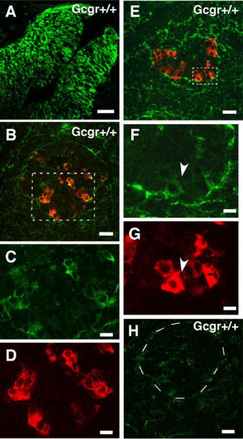
Fig. 1.
Expression of nestin by pancreatic endocrine cells of Gcgr+/+ embryos at e-11.5. A: Immunolocalization of nestin in radial glial cells of brain of e-11 mouse embryos. This photomicrograph documents the specificity of the nestin antibody from Chemicon. Similar results were obtained with the nestin antisera from Developmental Studies Hybrid-oma Bank (not shown). B: Pancreas of e-11 Gcgr+/+ embryo immuno-stained for glucagon (red) and nestin (green). Note the presence of glucagons+nestin+ cells (yellow). C,D: Area indicated with dotted lines is shown in higher magnification, which illustrates single label staining for nestin and glucagon respectively. Scale bars for B–D = 35 and 20 μm. E: Pancreas of e-11 Gcgr+/+ embryo immunostained for insulin (red) and nestin (green). F,G: Area indicated with dotted lines is magnified, which illustrates single label immunostaining for nestin and insulin, respectively. Cell indicated with arrowhead is positive for nestin and insulin. Scale bars for E–G = 35 and 10 μm. H: Photomicrograph of a section of pancreas of an e-11 Gcgr+/+ embryo incubated with a green fluorescent goat antimouse IgG (incubation with antibodies to nestin was omitted). Dotted circle indicates area containing an islet. This picture documents the absence of immunopositive nestin cells. Scale bar = 40 μm.

