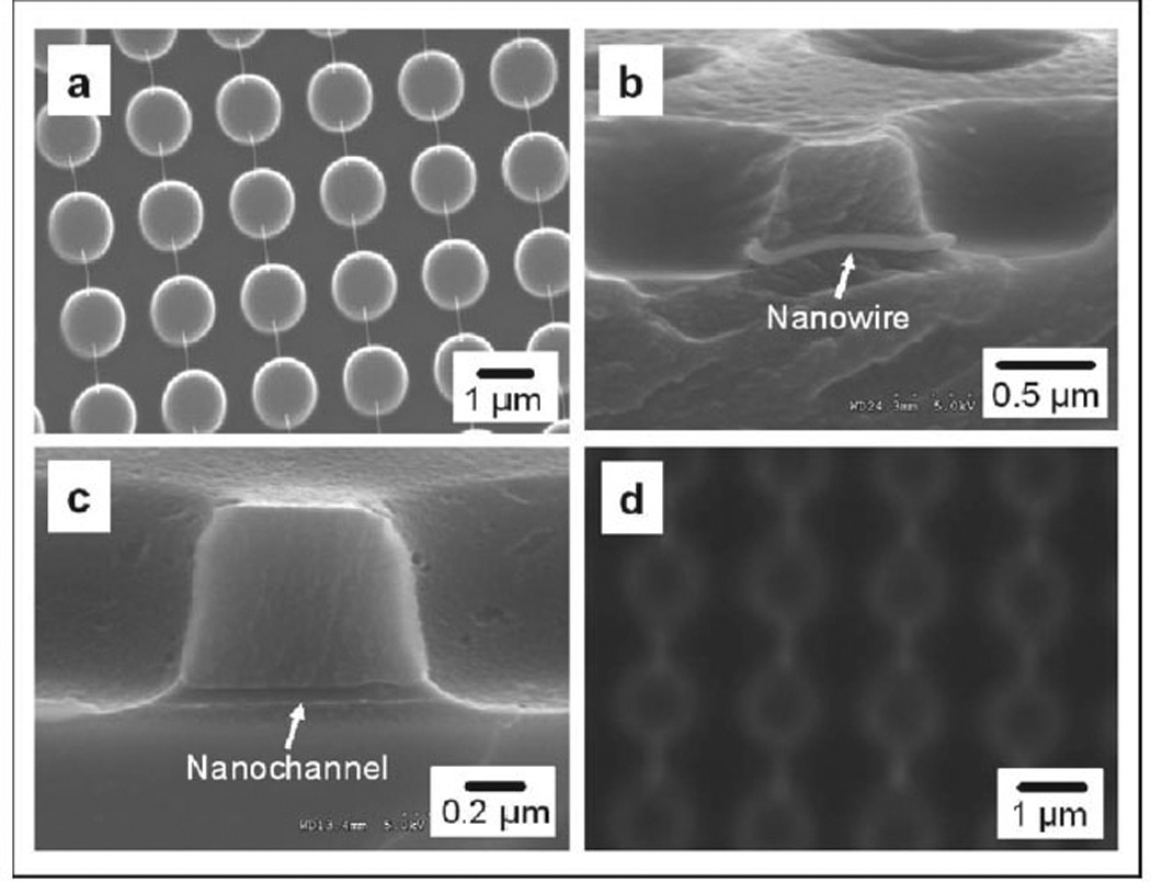
Figure 2.

Scanning electron microscopy (SEM) images of (a) suspended gold-coated DNA nanostrands between 1 µm wide micropillars, (b) a gold-coated DNA nanostrand embedded in the cured polymer, (c) side view of a nanochannel connecting two microwells, and (d) fluorescence micrograph of nanochannels and microwells filled with Rhodamine.
