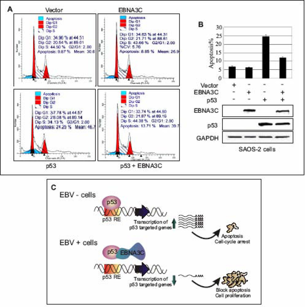
Figure 7.
EBNA3C blocks p53 induced apoptosis in p53 null cell line SAOS-2. (A–B) SAOS-2 (p53−/−) cells were transiently transfected either with vector control, or constructs expressing untagged wild-type EBNA3C and p53 alone, or p53 and EBNA3C together. After 48 h of transfection, cells were harvested, fixed and levels of apoptotic cells in each samples were analyzed by flow cytometry (Becton-Dickinson). Total 20,000 events were analyzed for each sample. Data were analyzed using the ModFIT model program (Verity Software House). The representative plot is a mean of three independent experiments. Error bar represents SD. EBNA3C (top panel) and p53 (middle panel) protein expression levels were judged by western blot. Equal protein loading was analyzed GAPDH (bottom panel) blot. (C) A schematic representation of p53 mediated transcriptional regulation in EBV negative (-) and positive (+) cells. In response to genotoxic stress, p53 achieves its antiproliferative properties through its action as a DNA-binding transcriptional activator, to induce expression of numerous downstream target genes, involved in cell-cycle arrest and apoptosis. In EBV positive cells, EBNA3C potentially inhibits p53 mediated transcriptional activity via forming a stable complex with p53. p53 RE: p53 responsive element.

