
Fig. 4.
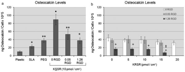
Effects of RGD, KRSR, and KSSR on osteocalcin levels in osteoblast-like MG63 cells. (a) Levels of osteocalcin increased on PLL-g-PEG surfaces compared with TCPS and SLA surfaces. The addition of 1.26 pmol/cm2 RGD decreased levels of osteocalcin. Addition of KSSR increased osteocalcin levels. (b) KRSR alone did not affect osteocalcin levels, although the combination of RGD and KRSR at high peptide density decreased osteocalcin levels. (a) *P<0 .05, Ti surfaces vs. plastic; **P<0.05, PEG surfaces vs. SLA; •P<0. 05. KSSR vs. PEG. (b) +P<0.05, RGD vs. KRSR alone (0 RGD) ; ‡P<0.0 5, KRSR vs. RGD alone (0 KRSR) .