
Fig. 6.
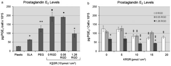
Effects of RGD, KRSR, and KSSR on PGE2 levels in osteoblast-like MG63 cells. (a) PGE2 levels were increased on PLL-g-PEG surfaces vs. TCPS and SLA surfaces. Addition of KSSR further increased levels of PGE2. (b) Addition of 1.26 pmol/cm2 RGD reduced levels of PGE2, as did addition of KRSR. Combining RGD and KRSR caused further inhibition of PGE2, especially at high surface peptide densities. (a) *<P0.05, Ti surfaces vs. plastic;**P<0.05, PEG surfaces vs. SLA. •P< 0.05, KSSR vs. PEG. (b) +P< 0.05, RGD vs. KRSR alone (0 RGD) ; ‡P<0.05, KRSR vs. RGD alone (0 KRSR).