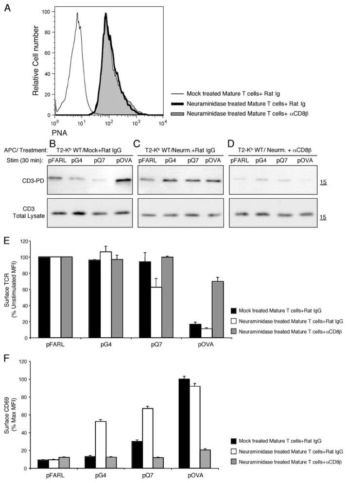
FIGURE 6.
Surface desialylation of mature T cells re-establishes preselection sensitivity for cognate ligand recognition and CD3Δc induction. A, Surface PNA staining (MFI) of CD8+ Kb WT/pOVA tetramer plus mature T cells from OT-I β2m+/+ lymph nodes that were mock and rat IgG-treated (B), neuraminidase and rat IgG-treated (C), or neuraminidase and anti-CD8β-treated (D). These T cells were cocultured with T2-Kb WT APC preloaded with pFARL, pQ7, pQ4H7, or pOVA (2 μM, 30 min, 37°C). The induction of CD3Δc was detected using the CD3-PD assay (top). Total CD3ζ content in the post-nuclear lysates is also shown (bottom). TCR down-regulation (E) and CD69 up-regulation (F) were analyzed by flow cytometry for the experiments shown in B–D. Peptides are listed on the x-axis, whereas surface TCR expression (E) is represented on the y-axis as a percentage of the expression level gated on Thy1+ CD4+CD8+ DP thymocytes cocultured with the null peptide pFARL. F, CD69 expression is represented on the y-axis as a percentage of the maximum induction on Thy1+ CD4+CD8+ DP thymocytes cocultured with peptide pOVA.

