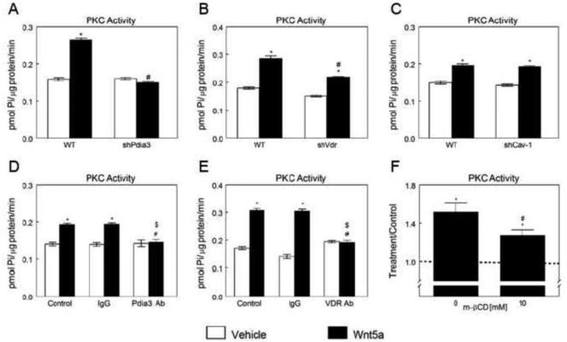
Figure 2.
Effects of Pdia3, Vdr and Cav-1 silencing or blocking and β-CD on Wnt5a-stimulated activation of PKC in MC3T3-E1 cells. Wnt5a-induced PKC activation in wild-type (WT), shPdia3 (A), shVdr (B) and shCav-1 (C) MC3T3-E1 cells. *p<0.05, treatment versus control; #p<0.05, versus Wnt5a treated WT. Effect of Pdia3 blocking (D) or VDR blocking (E) on Wnt5a-induced PKC activation. *p<0.05, treatment versus control; #p<0.05, versus Wnt5a treated control group; $p<0.05, versus Wnt5a treated IgG group. (F) Effect of β-CD on Wnt5ainducedPKC activation. Treatment over control ratios were calculated for each parameter. The dashed line represents the value for the control cultures, which was set to 1. *p<0.05, treatment versus control.

