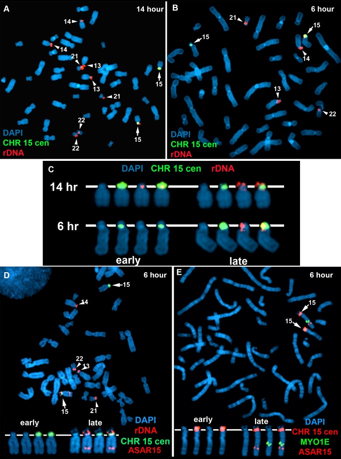
Figure 7. Coordinated random asynchronous replication on chromosome 15.
A–C) ReTiSH assay on rDNA loci in PBLs. PBLs were labeled with BrdU for 14 (A) or 6 (B) hours, arrested in metaphase, and subjected to ReTiSH using an 18S rDNA probe (red). The chromosome 15 s were identified using a centromeric probe (green), and the chromosomal DNA was detected with DAPI. A and B) The DAPI images of the chromosomes were inverted and the banding patterns were used to identify all of the ReTiSH positive chromosomes. The arrows mark the chromosome 15 s, and the arrowheads mark the other four chromosomes containing rDNA clusters (13, 14, 21, and 22). C) The ReTiSH signals for the rDNA (red) and chromosome 15 centromeric (green) probes from the 14 (panel A) and 6 (panel B) hour time points are shown. The early and late replicating chromosome 15 s are indicated for the 6 hour time point. D) ReTiSH assay using an ASAR15 BAC (CTD-2299E17; red), an rDNA probe (red), and a chromosome 15 centromeric probe (green). The ASAR15 and rDNA probes show hybridization signals to the same chromosome 15 homolog at the 6 hour time point. E) ReTiSH assay using an ASAR15 BAC (CTD-2299E17; red), a MYO1E BAC (RP11-1089J12; green) and a chromosome 15 centromeric probe (red). The ASAR15 BAC and the MYO1E BACs show hybridization signals to the same chromosome 15 homolog at the 6 hour time point. D and E) The early and late replicating chromosome 15 s are indicated.

