
Figure 1.
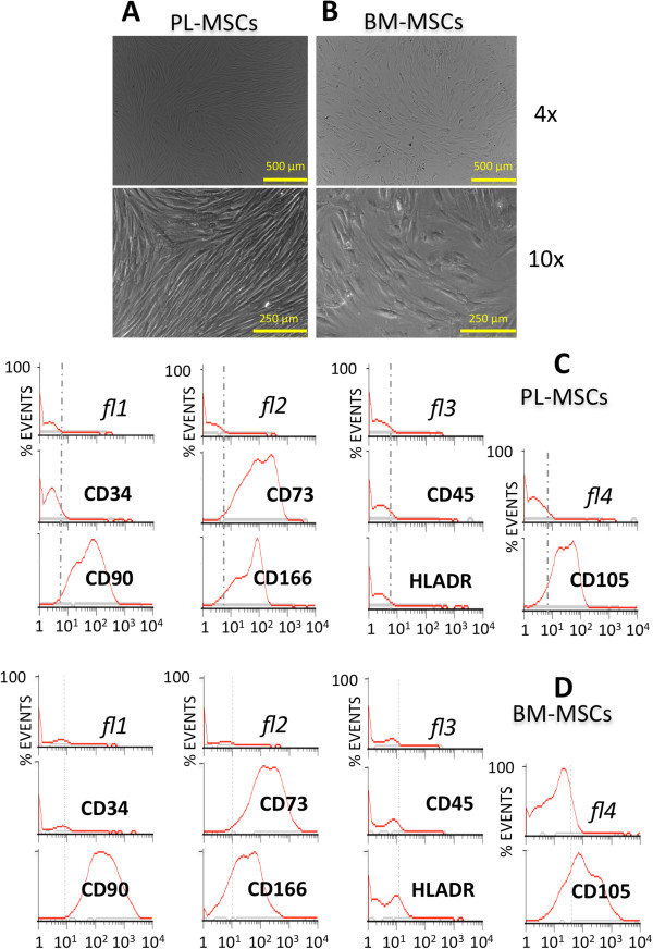
Characterization of MSCs primary cultures: microscope and flow cytometry. Microscope photographs of human MSCs in culture isolated from PL (A) and BM (B): phase contrast micrographs of passage three cultures seen at two amplifications. Flow cytometry histograms of standard immunophenotype markers (CD34, CD73, CD45, CD90, CD166, HLADR, CD105) tested in isolated PL (C) and BM (D) MSCs.