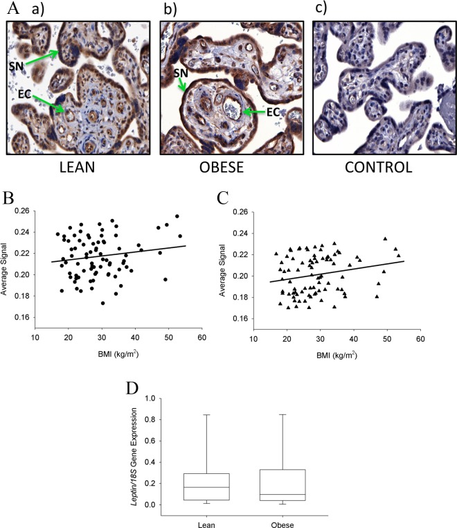
Figure 2.
Immunostaining of leptin in placental tissue. A, Examples of placental sections immunostained for leptin from a lean (a) and obese woman (b), and negative control (c). Leptin immunostaining was localized to the syncytiotrophoblast (SN) and the villous vascular endothelial cells (EC). B, The quantitation of immunostaining in the syncytiotrophoblast showed a nonsignificant trend for increasing leptin levels with increasing maternal body mass index (BMI; P = .156). C, The quantitation in the villous vascular endothelial cells was significantly and linearly increased with increasing maternal BMI (P = .046). D, The expression of leptin messenger RNA (mRNA) in the 2 extreme groups (lean and obese women) in the placental villous trophoblast showed no significant differences (P = .5). Original magnification 400× for all sections.

