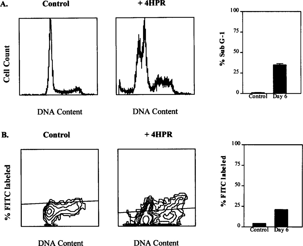
Fig. 3.
A, effects of fenretinide (3 µM) on cell cycle kinetics of D54 cells. Control and fenretinide-treated D54 cells were harvested on days 1, 3, and 6 and studied by flow cytometric analysis. Fenretinide-treated cells showed no changes in cell cycle distribution compared with control cells. A sub-G1 population indicating cells with fragmented DNA (<2N) was seen in fenretinide-treated cells by day 3 and increased to a maximum by day 6. Bars, SE. B, D54 cells treated with 3 µM fenretinide were harvested on days 3 and 6 with untreated control cells and studied by TUNEL assay using the APODIRECT kit. FITC-labeled cells representing the apoptotic subpopulation undergoing DNA fragmentation were quantified. Apoptosis was seen maximally by day 6 and occurred in a cell cycle-independent manner as shown.

