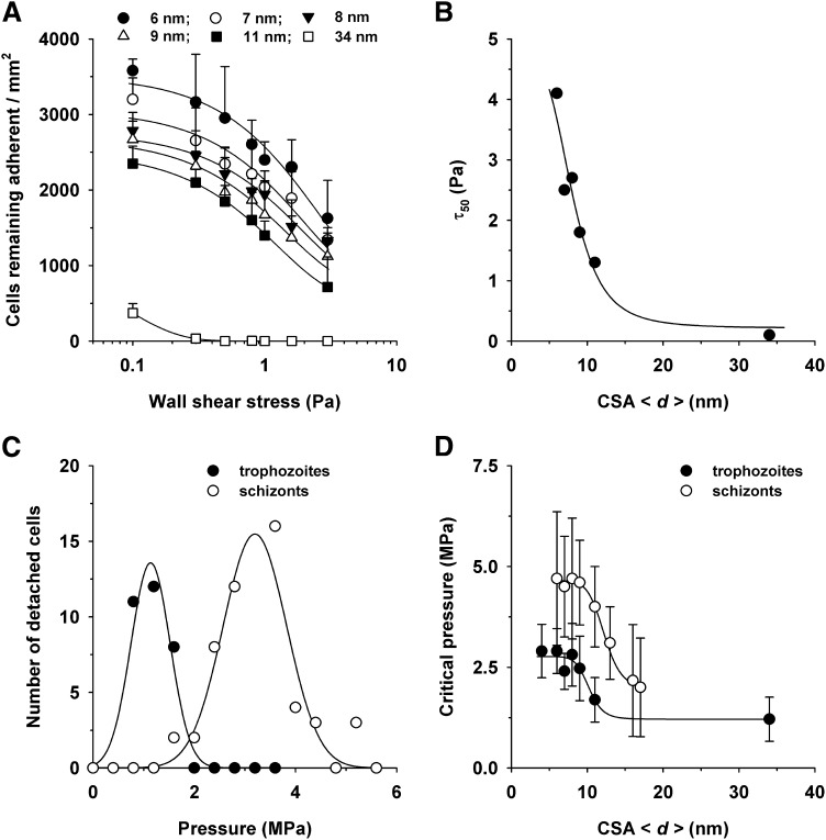
Figure 3.
Effect of the intermolecular CSA distance on the detachment of erythrocytes infected with FCR3CSA. (A) Cells (4 × 10⁷ mL−1) were allowed to settle on the CSA-functionalized membranes for 60 minutes under controlled atmospheric conditions before gradually increasing wall shear stresses (ranging from 0.1 to 5 Pa) were applied. The number of adherent cells were subsequently determined for each hydrodynamic condition and analyzed as a function of the wall shear stress. The mean ± SD of at least 4 biological replicates are shown. The data points were fitted using an exponential decay function. (B) The wall shear stress at which 50% of the cells detached (τ50) was determined for each hydrodynamic condition and analyzed as a function of the CSA distance. The data were fitted using a Hill function. (C) Erythrocytes infected with FCR3CSA at the trophozoite and schizont stages were allowed to adhere on a functionalized membrane with an intermolecular CSA distance of 6 nm for 60 minutes under controlled atmospheric conditions. Noncorpuscular ultrasonic pressure waves ranging from 0.3 to 5.6 MPa were applied, and the number of detachment events was observed as a function of the hydrodynamic pressure. (D) The critical pressures at which 95% of the adherent cells detached are shown as a function of the intermolecular CSA distance. The data points were fitted using a Hill function. The means ± SD of at least 3 biological replicates are shown.

