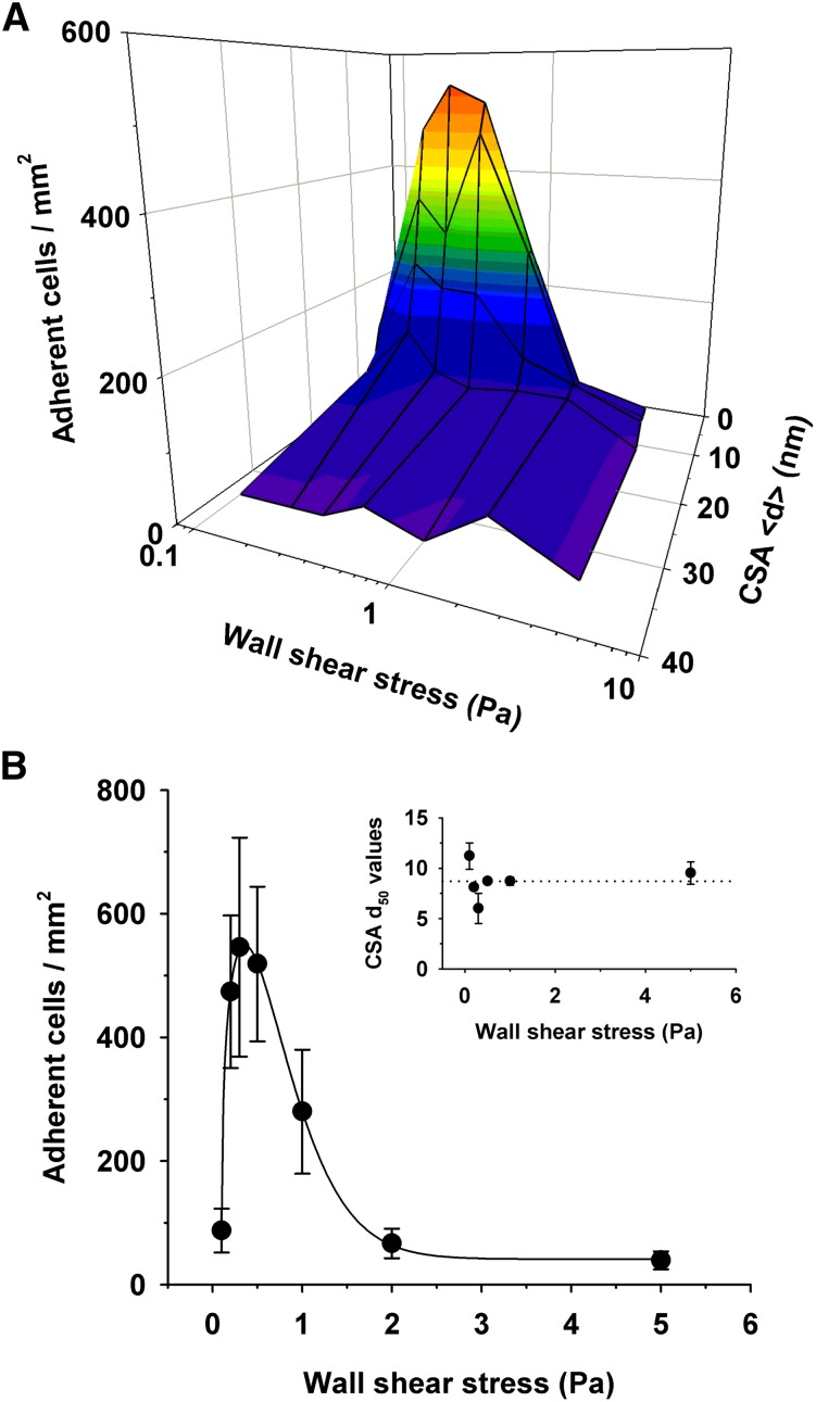
Figure 4.
Effect of the intermolecular CSA distance on adhesion of erythrocytes infected with FCR3CSA in flow. (A) Parasitized erythrocytes at the trophozoite stage (1 × 10⁶) were streamed through microfluidic chambers containing different CSA arrangements at the hydrodynamic conditions indicated. The number of adhering cells per square millimeter is shown as a function of the intermolecular CSA distance and the wall shear stress. (B) The number of cells adhering to a functionalized membrane with a CSA distance of 7 nm at different wall shear stresses. Inset: intermolecular CSA distance at which 50% of the cells adhere (d50) as a function of the wall shear stress. The d50 values were obtained by curve fitting the data points for each hydrodynamic condition. The means ± SD of at least 3 biological replicates are shown.

