Figure 5.
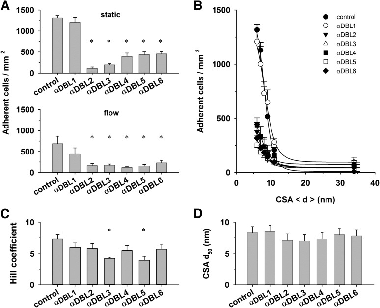
Effect of DBL domain–specific antibodies on cytoadhesion. (A) Erythrocytes (4 × 10⁷ mL−1) infected with FCR3CSA were incubated with preadsorbed antibodies directed against the DBL domains indicated (dilution 1:40). Upper panel: cells were subsequently allowed to settle on CSA functionalized membranes (6-nm spacing) for 30 minutes before unbound cells were washed off and the number of adherent cells determined. Lower panel: the adhesion behavior was determined in laminar flow at a wall shear stress of 0.2 Pa. The means ± SD of 4 biological replicates are shown. All assays were performed in parallel. (B) Number of adherent cells as a function of the CSA distance under static conditions. The curves presented in panel B were fitted using a Hill function, and the values for the Hill coefficient (C) and the CSA distance that supports half-maximal adhesion (d50) (D) were derived and analyzed as a function of the DBL domain–specific antibodies. *P < .01.

