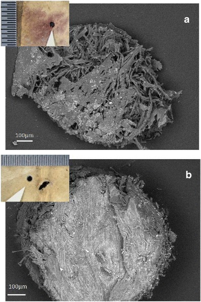
Fig. 2.

Core samples taken from purple stained and degraded (a) and healthy/control areas (b) of folio 68v. a SEM low magnification image of the core taken from a stained and degraded area (location B in b), and obtained at variable pressure (50 Pa) with a backscattered electron detector on uncoated material. b SEM low magnification image of the core obtained from a healthy/control area (location C in Fig. 1b). Inset in both images are photomicrographs taken at the Walters Art Museum of the tiny holes left in the parchment after the core samples were removed (Images taken at the Walters Art Museum are copyright of the owner of the Archimedes Palimpsest)
