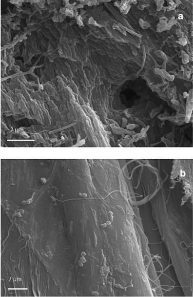
Fig. 5.

Samples taken from a stained/degraded area (a) and a healthy/control area of folio 68v (b). The pictures were obtained with high-vacuum secondary electron SEM imaging on gold-sputtered core samples; scale, 2 μm. A profound structural damage consisting of holes, cracks, and fissures was documented by HV-SEM imaging on both the flesh and hair sides of the first sample (a). b Shows a collagen fiber from the healthy parchment core sample; the surface is smooth and compact, although a few bacterial cells, appearing in chains, were observed adhering to the surface of the fiber
