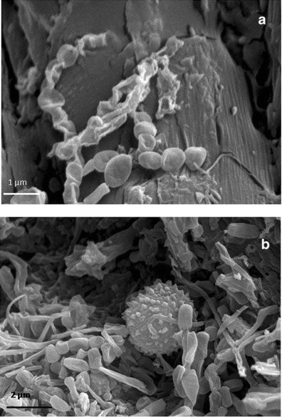
Fig. 6.

HV-SEM images of gold-sputtered core samples of parchment taken from folio 68v. a Taken from the core sample corresponding to the “healthy” area; it shows the bacterial structure at the initial formation of a spore chain. It represents an initial attack to the collagen fiber, which is still visible and integer on the backward; bacterial cells (less than 1 μm in diameter) form chains that are morphologically consistent with Actinomycetales taxon; the filaments appear branched and are fragmenting into spores; scale, 1 μm. b Taken from the core sample corresponding to the “purple/damaged” area; it shows chains of bacterial spores and filaments and a single fungal conidia, globose, and with echinated ornamentation; the collagen fiber is no longer distinguishable; scale, 2 μm
