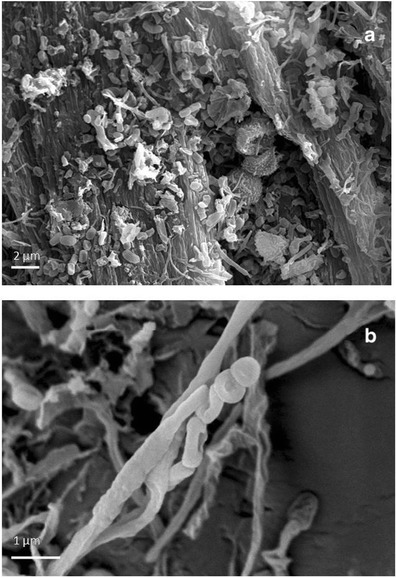
Fig. 7.

HV-SEM images of gold-sputtered core samples of stained/degraded parchment from two different folios. a Chains of bacterial spores and filaments and a chain of fungal conidia, globose, and with echinated ornamentation; the fungal conidia appear covered with bacterial filaments. In the background, the degraded collagen fibers are also visible; scale 2 μm. b Aerial bacterial filaments appear coarsely wrinkled and branched, fragmenting into spores; scale, 1 μm
