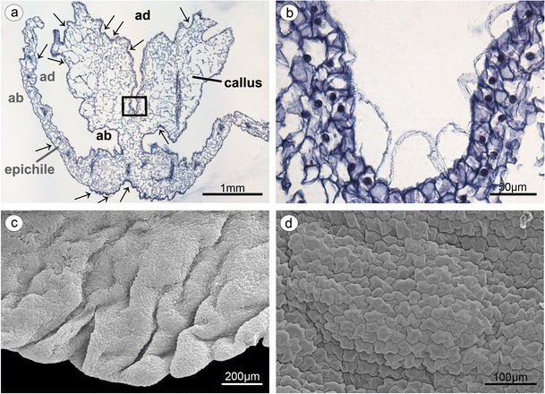
Fig. 7.

a Epichile cross-section of the epichile (tepal and callus) with distended cuticle (arrows) on adaxial surface (ad) and abaxial surface (ab) (paraffin method). b Magnification of a, central groove with distended cuticle (paraffin method). c Strongly undulate margins of epichile (SEM). d Magnification of c, the epichile surface built by polygonal cells with U-shaped cell walls (SEM)
