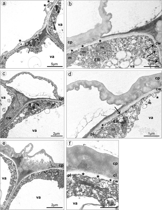
Fig. 8.

Ultrastructure of callus cells. a The swelled and almost ruptured cuticle (asterisks) caused by pressure of gathered substances, the globules noted beneath the cuticle (arrows). b Spherical mitochondria located near the plasmalemma (asterisks), profiles of ER in contact with plasmalemma (arrows). c The bulges of distended cuticle on the surface upon the border of neighboring cells and distended further on the whole cell surface, the cell volume occupied by a central vacuole, parietal cytoplasm. d Profiles of ER in contact with plasmalemma (arrow), vesicles building into plasmalemma (asterisks), the cuticle consisted of cuticle proper and reticulate cuticle layer. e The swelled cuticle on the surface upon the border of neighboring cells. f Vesicles building into plasmalemma (asterisks), dictyosome (c cuticle, cl cuticle layer, cp cuticle proper, cw cell wall, d dictyosome, ER endoplasmic reticulum, m mitochondrion, pl plasmalemma, va vacuole)
