
Figure 7. Effects of M645C subunits on the voltage dependence of hERG1 inactivation.
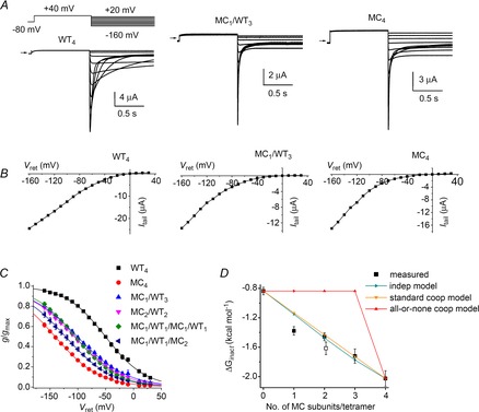
A, representative current traces for WT4, MC1/WT3 and MC4 concatenated tetramers recorded during pulses to indicated voltages. Oocytes were bathed in 96K4Na extracellular solution. B, fully activated Itail–Vret relationships for currents shown in (A). C, voltage dependence of inactivation for indicated concatenated tetramers. Values of V0.5 and z determined from fitting data to Boltzmann function are summarized in Table 1. D, ΔGinact calculated from data (squares, n = 4–6) plotted as a function of the number of mutant M645C subunits contained in a concatenated tetramer together with ΔGinact predicted by the indicated independent and cooperative models. Open square represents MC1/WT1/MC1/WT1 channels.