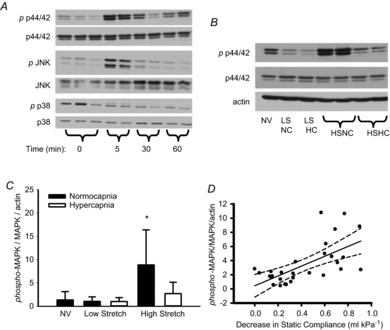
Figure 2. MAPK activation in mouse model of VILI.

A, time course of MAPK activation during high stretch ventilation of mouse lung. B, representative Western blot demonstrates phosphorylation of p44/42 MAPK at 3 h in mechanically ventilated mouse lungs. C, p44/42 MAPK is activated by high VT only under normocapnia. *P < 0.05 vs. all other groups, n = 5 NV and LS groups, n = 10 HS groups. D, linear regression analysis indicating correlation between decrease in compliance and activation of p44/42 MAPK in ventilated mice. r2 = 0.406, P < 0.001, n = 30. HC, hypercapnia; HS high stretch; LS, low stretch; MAPK, mitogen-activated protein kinase; NC, normocapnia; NV, non-ventilated.
