
Figure 4. EGFR activation in mouse model of VILI.
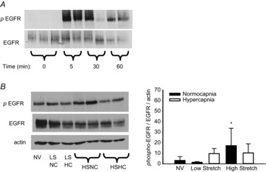
A, time course of EGFR activation during high stretch ventilation of mouse lung. B, following 3 h ventilation, EGFR remains significantly activated only in HSNC. †P < 0.05 vs. NV and LSNC, no other differences; n = 5 NV and LS groups, n = 10 HS groups. EGFR, epidermal growth factor receptor; HC, hypercapnia; HS high stretch; LS, low stretch; NC, normocapnia; NV, non-ventilated.