
Figure 5. Mitochondrial dysfunction is established early during HU in soleus muscle.
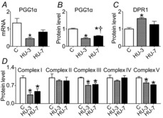
A, quantification of mRNA levels of PGC-1α by real-time PCR. B, quantification of protein levels of PGC-1α by Western blot. C, quantification of protein levels of DRP1 involved in fission machinery by Western blot. D, quantification of protein levels of mitochondrial complexes by Western blot. C, control; HU-3, 3 days of hindlimb unloading; HU-7, 7 days of hindlimb unloading. *Significantly different from control (P < 0.05); †significantly different from HU-3 (P < 0.05). Data are presented as means ± SEM.