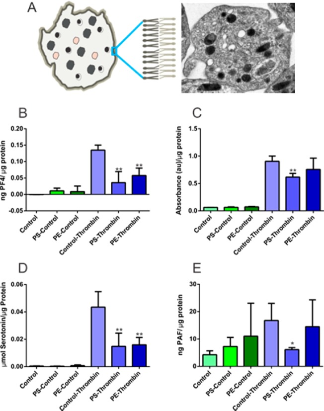
Figure 2.
Assessment of phospholipid effects on ensemble platelet granule secretion. (A) Transmission electron micrograph of a platelet and a representative figure illustrating three different platelet granule types; δ-granule, bull’s eye shape; lysosome, pink shape; and α-granule, uniformly filled irregular shapes. Phospholipids are asymmetrically distributed in the platelet membrane. (B) PF4 release from α-granules decreased with enrichment of each of the phospholipids studied. (C) Lysosomal release decreased with PS enrichment. (D) δ-granule secretion was also suppressed upon incubation with phospholipids. (E) PAF secretion was suppressed upon PS enrichment. *p < 0.05 and **p < 0.01 compared to thrombin-stimulated control platelets.

