
Figure 2.
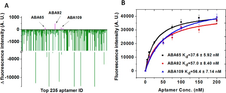
Identification of three aptamers that form binding pairs with ABA1. (A) We used our aptamer array to identify aptamers that can bind Ang2 in complex with ABA1. Negative ΔF (green) indicates an aptamer feature on the array that binds the same site on Ang2 as ABA1, while non-negative ΔF (magenta) indicates aptamer features that bind a different site on Ang2 than ABA1. (B) We identified three candidate aptamers (ABA65, ABA92 and ABA109) that potentially form binding pairs with ABA1, and measured their Kds using a bead-based fluorescence assay. As expected, their affinities were moderately lower than that of ABA1. Fluorescence intensities are the average of triplicate measurements.