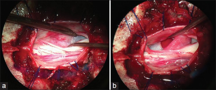
Figure 2.

Intraoperative photograph. (a) Here, the herniated spinal cord content is demonstrated (instrument tip to the right). (b) The dural defect (indicated by the instrument), the reduced spinal cord area, and the tapering of the spinal cord at the herniation level are shown
