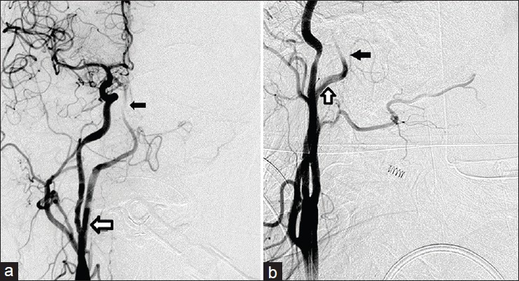
Figure 1.

Digital subtraction angiography, AP (a) and lateral view (b), revealing the mid-basilar occlusion and the origin of the PPHA from the cervical segment of the ICA. The occlusion is marked with a small black arrow. The PPHA is marked with a white arrow
