
Figure 1. Tumor cells induce T-cell senescence.
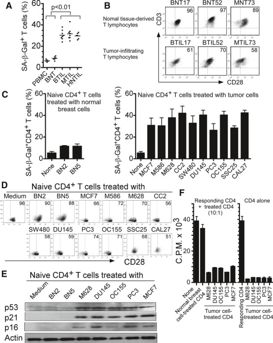
A, B Increased SA-β-Gal-positive T-cell populations (A) and decreased CD28 expression (B) existed in TILs, compared with those in normal tissue-derived lymphocytes. CD3+ T cells were purified from the cultured tissue-infiltrating T cells from freshly digested tumor or normal tissues by microbeads and SA-β-Gal staining or CD28 expression was determined. BNT: normal breast tissue-derived T cells; BTIL, MTIL, and HNTIL: TIL obtained from breast cancer, melanoma, and head and neck cancers, respectively.
C Tumor cell treatment significantly increased SA-β-Gal-positive T-cell populations in co-cultured naïve CD4+ T cells. However, naïve CD4+ T cells cultured in medium only or co-cultured with normal breast cells had little SA-β-Gal expression. Anti-CD3-activated naïve CD4+ T cells were co-cultured with different types of tumor cells or normal breast cells at ratio of 1:1 for 1 day. The treated CD4+ T cells were then separated and stained with SA-β-Gal staining reagents after culture for additional 3 days. Cell lines included: breast cancer (MCF7), melanoma (M586 and M628), colon cancer (SW480 and CC2), ovarian cancer (OC155), prostate cancer (DU145 and PC3), squamous cancers (SSC25 and CAL27), and normal breast cells (BN2 and BN5).
D Decreased CD28 expression in naïve CD4+ T cells after culture with different types of tumor cell lines but not normal breast cells. Cell treatment and procedure were the same as in (C). CD28 expression in treated naïve CD4+ T cells were analyzed by flow cytometry.
E Increased expression of cell cycle regulatory molecules p53, p21, and p16 in naïve T cells treated with tumor cells. Cell treatment and procedure were the same as in (C). Co-cultured naïve CD4+ T cells were purified and cell lysates were prepared for Western blot analyses.
F Suppressive function of tumor-induced senescent T cells. Senescent CD4+ T cells induced by different types of tumor cell lines strongly inhibited the proliferation of responding CD4+ T cells. In contrast, naïve T cells treated or untreated with normal breast cells did not affect the proliferation of responding CD4+ T cells (left panel). Furthermore, senescent CD4+ T cells induced by different types of tumor cell lines did not proliferate themselves (right panel). Cell treatment and procedure were the same as in (C). Treated CD4+ T cells were purified, and the suppressive activity on responding CD4+ T-cell proliferation was evaluated using [3H]-thymidine incorporation assays.
Data information: Data are representative of average of three independent experiments ± SD.