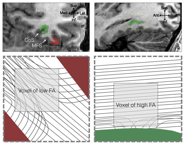
Figure 8. Local axonal environment and its relationship to tractography and FA.
Upper left: A coronal section of the VTC. Upper right: A sagittal section taken at the fundus of the collateral sulcus. Red: Face selective cortex on the lateral fusiform gyrus; Green: place selective cortex on the collateral sulcus (CoS). Bottom left: an enlarged view of the dotted rectangle from the coronal section. Voxels within a gyrus will contain axons in multiple directions, lowering FA and making tractography near the cortical surface difficult. Increases in local connectivity to the crown of the gyrus will likely result in higher FA. Bottom right: enlarged view of the dotted rectangle from the sagittal slice. The fundus of the collateral sulcus is exposed to longitudinal running axons. Because these tracts are closer to cortex, increased local connectivity to cortex would lower FA.

