

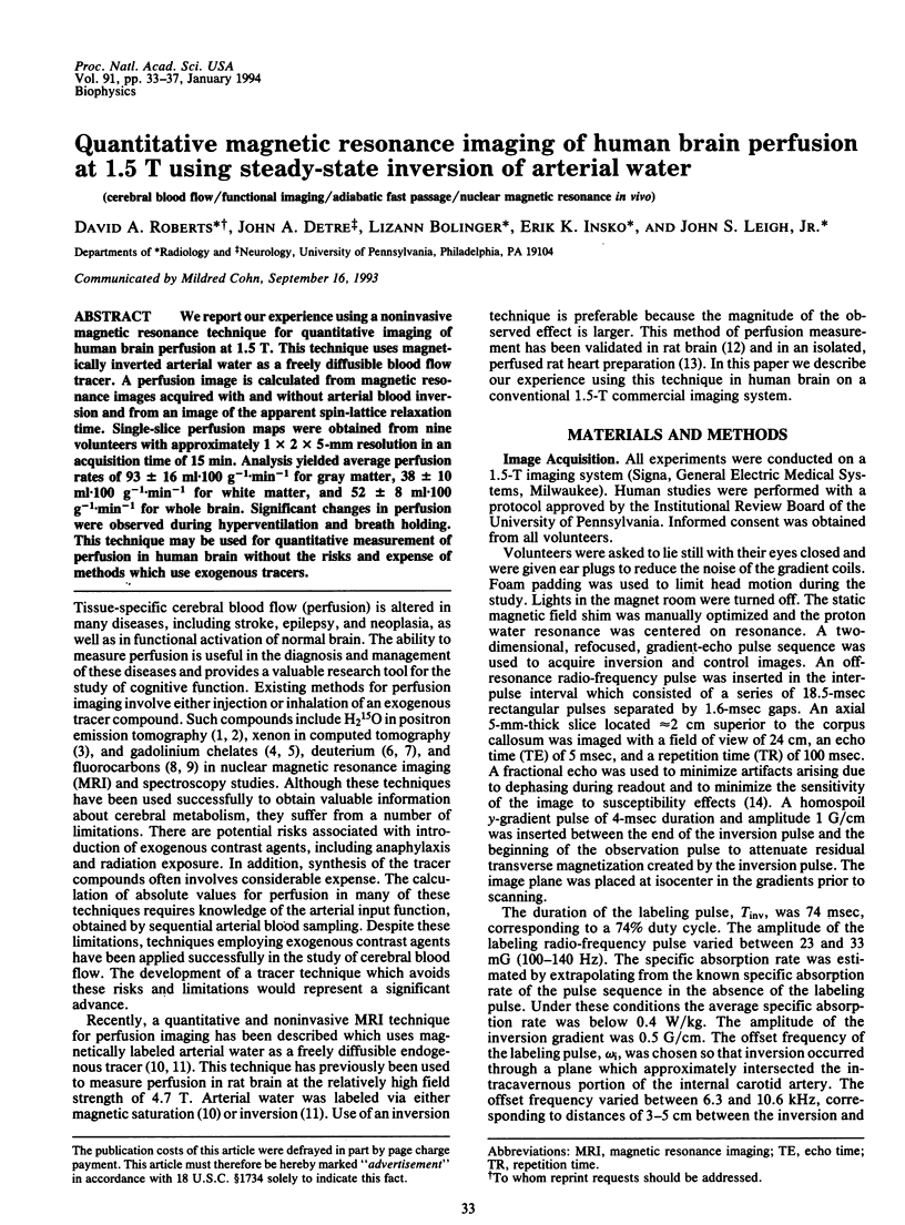
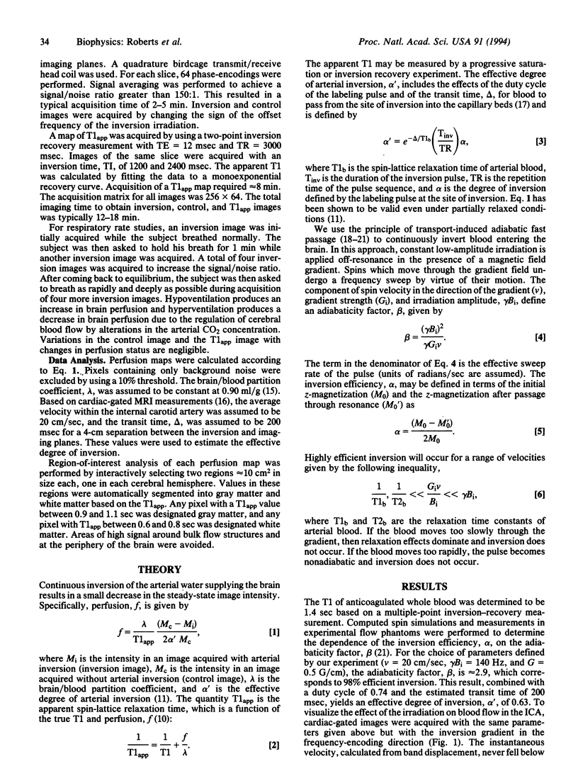
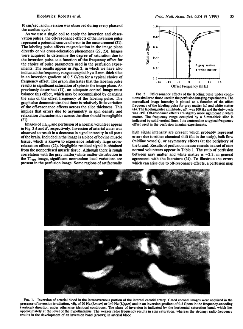
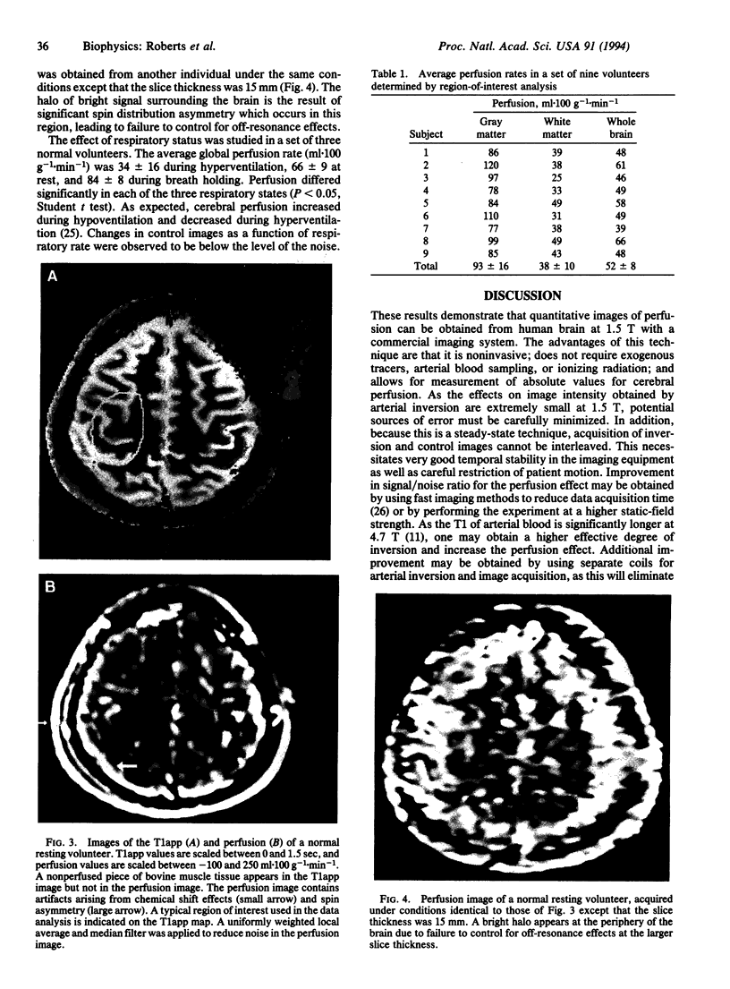
Abstract
We report our experience using a noninvasive magnetic resonance technique for quantitative imaging of human brain perfusion at 1.5 T. This technique uses magnetically inverted arterial water as a freely diffusible blood flow tracer. A perfusion image is calculated from magnetic resonance images acquired with and without arterial blood inversion and from an image of the apparent spin-lattice relaxation time. Single-slice perfusion maps were obtained from nine volunteers with approximately 1 x 2 x 5-mm resolution in an acquisition time of 15 min. Analysis yielded average perfusion rates of 93 +/- 16 ml.100 g-1.min-1 for gray matter, 38 +/- 10 ml.100 g-1.min-1 for white matter, and 52 +/- 8 ml.100 g-1.min-1 for whole brain. Significant changes in perfusion were observed during hyperventilation and breath holding. This technique may be used for quantitative measurement of perfusion in human brain without the risks and expense of methods which use exogenous tracers.
Full text
PDF
Images in this article
Selected References
These references are in PubMed. This may not be the complete list of references from this article.
- Ackerman J. J., Ewy C. S., Becker N. N., Shalwitz R. A. Deuterium nuclear magnetic resonance measurements of blood flow and tissue perfusion employing 2H2O as a freely diffusible tracer. Proc Natl Acad Sci U S A. 1987 Jun;84(12):4099–4102. doi: 10.1073/pnas.84.12.4099. [DOI] [PMC free article] [PubMed] [Google Scholar]
- Belliveau J. W., Kennedy D. N., Jr, McKinstry R. C., Buchbinder B. R., Weisskoff R. M., Cohen M. S., Vevea J. M., Brady T. J., Rosen B. R. Functional mapping of the human visual cortex by magnetic resonance imaging. Science. 1991 Nov 1;254(5032):716–719. doi: 10.1126/science.1948051. [DOI] [PubMed] [Google Scholar]
- Detre J. A., Eskey C. J., Koretsky A. P. Measurement of cerebral blood flow in rat brain by 19F-NMR detection of trifluoromethane washout. Magn Reson Med. 1990 Jul;15(1):45–57. doi: 10.1002/mrm.1910150106. [DOI] [PubMed] [Google Scholar]
- Detre J. A., Subramanian V. H., Mitchell M. D., Smith D. S., Kobayashi A., Zaman A., Leigh J. S., Jr Measurement of regional cerebral blood flow in cat brain using intracarotid 2H2O and 2H NMR imaging. Magn Reson Med. 1990 May;14(2):389–395. doi: 10.1002/mrm.1910140223. [DOI] [PubMed] [Google Scholar]
- Dixon W. T., Du L. N., Faul D. D., Gado M., Rossnick S. Projection angiograms of blood labeled by adiabatic fast passage. Magn Reson Med. 1986 Jun;3(3):454–462. doi: 10.1002/mrm.1910030311. [DOI] [PubMed] [Google Scholar]
- Eleff S. M., Schnall M. D., Ligetti L., Osbakken M., Subramanian V. H., Chance B., Leigh J. S., Jr Concurrent measurements of cerebral blood flow, sodium, lactate, and high-energy phosphate metabolism using 19F, 23Na, 1H, and 31P nuclear magnetic resonance spectroscopy. Magn Reson Med. 1988 Aug;7(4):412–424. doi: 10.1002/mrm.1910070404. [DOI] [PubMed] [Google Scholar]
- Firmin D. N., Klipstein R. H., Hounsfield G. L., Paley M. P., Longmore D. B. Echo-planar high-resolution flow velocity mapping. Magn Reson Med. 1989 Dec;12(3):316–327. doi: 10.1002/mrm.1910120304. [DOI] [PubMed] [Google Scholar]
- Frahm J., Merboldt K. D., Hänicke W. Functional MRI of human brain activation at high spatial resolution. Magn Reson Med. 1993 Jan;29(1):139–144. doi: 10.1002/mrm.1910290126. [DOI] [PubMed] [Google Scholar]
- Gur D., Good W. F., Wolfson S. K., Jr, Yonas H., Shabason L. In vivo mapping of local cerebral blood flow by xenon-enhanced computed tomography. Science. 1982 Mar 5;215(4537):1267–1268. doi: 10.1126/science.7058347. [DOI] [PubMed] [Google Scholar]
- Herscovitch P., Markham J., Raichle M. E. Brain blood flow measured with intravenous H2(15)O. I. Theory and error analysis. J Nucl Med. 1983 Sep;24(9):782–789. [PubMed] [Google Scholar]
- Herscovitch P., Raichle M. E. What is the correct value for the brain--blood partition coefficient for water? J Cereb Blood Flow Metab. 1985 Mar;5(1):65–69. doi: 10.1038/jcbfm.1985.9. [DOI] [PubMed] [Google Scholar]
- Kwong K. K., Belliveau J. W., Chesler D. A., Goldberg I. E., Weisskoff R. M., Poncelet B. P., Kennedy D. N., Hoppel B. E., Cohen M. S., Turner R. Dynamic magnetic resonance imaging of human brain activity during primary sensory stimulation. Proc Natl Acad Sci U S A. 1992 Jun 15;89(12):5675–5679. doi: 10.1073/pnas.89.12.5675. [DOI] [PMC free article] [PubMed] [Google Scholar]
- Lee H. K., Nalcioglu O., Moran P. R. Spatially resolved flow velocity measurements and projection angiography by adiabatic passage. Magn Reson Imaging. 1991;9(1):115–127. doi: 10.1016/0730-725x(91)90105-u. [DOI] [PubMed] [Google Scholar]
- Mansfield P., Maudsley A. A. Medical imaging by NMR. Br J Radiol. 1977 Mar;50(591):188–194. doi: 10.1259/0007-1285-50-591-188. [DOI] [PubMed] [Google Scholar]
- Ogawa S., Lee T. M., Nayak A. S., Glynn P. Oxygenation-sensitive contrast in magnetic resonance image of rodent brain at high magnetic fields. Magn Reson Med. 1990 Apr;14(1):68–78. doi: 10.1002/mrm.1910140108. [DOI] [PubMed] [Google Scholar]
- Roberts D. A., Bolinger L., Detre J. A., Insko E. K., Bergey P., Leigh J. S., Jr Continuous inversion angiography. Magn Reson Med. 1993 May;29(5):631–636. doi: 10.1002/mrm.1910290508. [DOI] [PubMed] [Google Scholar]
- Rosen B. R., Belliveau J. W., Vevea J. M., Brady T. J. Perfusion imaging with NMR contrast agents. Magn Reson Med. 1990 May;14(2):249–265. doi: 10.1002/mrm.1910140211. [DOI] [PubMed] [Google Scholar]
- Schuierer G., Ladebeck R., Barfuss H., Hentschel D., Huk W. J. Sodium-23 imaging of supratentorial lesions at 4.0 T. Magn Reson Med. 1991 Nov;22(1):1–9. doi: 10.1002/mrm.1910220102. [DOI] [PubMed] [Google Scholar]
- Ter-Pogossian M. M., Eichling J. O., Davis D. O., Welch M. J., Metzger J. M. The determination of regional cerebral blood flow by means of water labeled with radioactive oxygen 15. Radiology. 1969 Jul;93(1):31–40. doi: 10.1148/93.1.31. [DOI] [PubMed] [Google Scholar]
- Williams D. S., Detre J. A., Leigh J. S., Koretsky A. P. Magnetic resonance imaging of perfusion using spin inversion of arterial water. Proc Natl Acad Sci U S A. 1992 Jan 1;89(1):212–216. doi: 10.1073/pnas.89.1.212. [DOI] [PMC free article] [PubMed] [Google Scholar]
- Wolff S. D., Balaban R. S. Magnetization transfer contrast (MTC) and tissue water proton relaxation in vivo. Magn Reson Med. 1989 Apr;10(1):135–144. doi: 10.1002/mrm.1910100113. [DOI] [PubMed] [Google Scholar]
- Zhang W., Williams D. S., Detre J. A., Koretsky A. P. Measurement of brain perfusion by volume-localized NMR spectroscopy using inversion of arterial water spins: accounting for transit time and cross-relaxation. Magn Reson Med. 1992 Jun;25(2):362–371. doi: 10.1002/mrm.1910250216. [DOI] [PubMed] [Google Scholar]
- Zhang W., Williams D. S., Koretsky A. P. Measurement of rat brain perfusion by NMR using spin labeling of arterial water: in vivo determination of the degree of spin labeling. Magn Reson Med. 1993 Mar;29(3):416–421. doi: 10.1002/mrm.1910290323. [DOI] [PubMed] [Google Scholar]