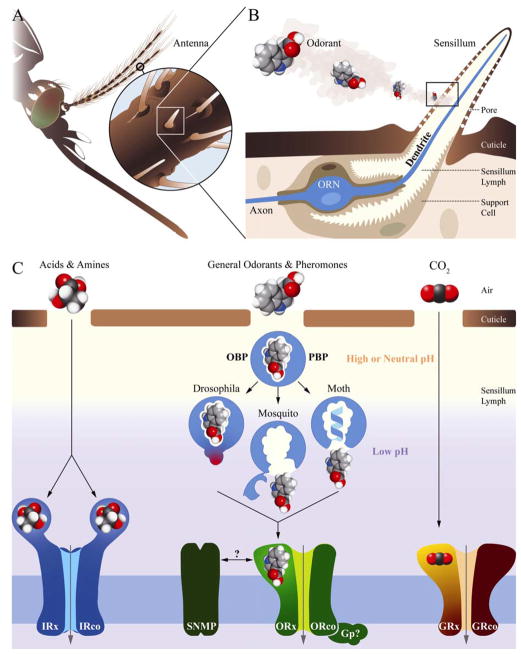
Figure 1.
(A) Insect chemosensory organs and molecular models in signal transduction. Insect chemosensory appendages such as the antennae, maxillary palps and labials are covered by sensilla. (B) An olfactory sensillum housing support cells and an ORN (blue); odorants encounter the ORN dendrite across sensillum lymph after penetration via cuticular pores. (C) Distinct classes of odorants activate specific groups of chemoreceptors and other components with diverse mechanistic models: Drosophila may use the bound OBP to activate the receptors. Moth pheromone-binding proteins (PBPs) eject their odorant load through a pH-induced conformational change including the formation of an α-helix that occupies the binding pocket. Mosquito odorant-binding proteins (OBPs) also eject their ligands through a pH-induced conformation change including the formation of a ‘lid’.

