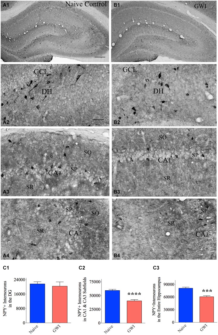
Figure 2.
Exposure to Gulf War illness related (GWIR) chemicals and stress causes loss of the neuropeptide Y-positive (NPY+) interneurons in the hippocampus. Panels (A1) and (B1) illustrate the distribution of NPY+ interneurons in different subfields of the hippocampus of an age-matched naive control rat (A1) and a rat that was exposed to GWIR-chemicals and stress 3 months earlier (B1). Panels (A2–A4) show magnified views of the dentate gyrus (DG), the CA1 subfield and the CA3 subfield from the Panel (A1). Panels (B2–B4) show magnified views of the DG, the CA1 subfield and the CA3 subfield from the Panel (B1). Reduced density of NPY+ interneuron cell bodies is apparent in the CA1 and CA3 subfields of a GWI rat (B3, B4), in comparison to respective regions from a naive control rat (A3, A4). Scale bar (A1) and (B1) = 500 μm; (A2–A4) and (B2–B4) = 100 μm. Bar charts in (C1–C3) compare numbers of NPY+ interneurons in the DG (C1), CA1 and CA3 subfields (C2) and the entire hippocampus (C3) between naive control rats and GWI-rats. ***p < 0.001; ****p < 0.0001 (two tailed, unpaired Student’s t-test). DH, dentate hilus; GCL, granule cell layer; SO, stratum oriens, SP, stratum pyramidale; SR, stratum radiatum.

