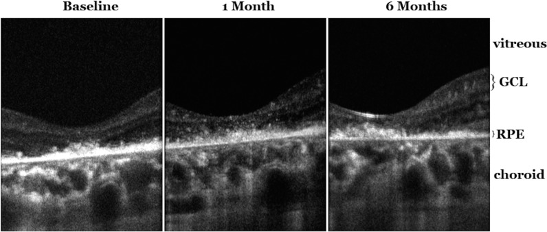
Figure 5.
Adaptive optics optical coherence tomography (AO-OCT) imaging of the central macula of the study eye of subject with Stargardt's disease. Left: B-scan cross-sectional images of the central macula at study enrollment showing the irregular hyperreflective retinal pigment epithelial layer with overlying photoreceptor inner segment–outer segment layer not well visualized. Middle: Cross-sectional B-scan images of the central macula 1 month after intravitreal CD34+ cell injection showing new multiple hyperreflective (white) deposits within the retinal layers. Right: Cross-sectional B-scan image taken 6 months after CD34+ cell injection showing less prominent hyperreflective deposits within the retinal layers and a hint of an increase in hyperreflectivity of the irregular retinal pigment epithelial layer centrally under the fovea. GCL, ganglion cell layer; RPE, retinal pigment epithelium.

