
Figure 2.
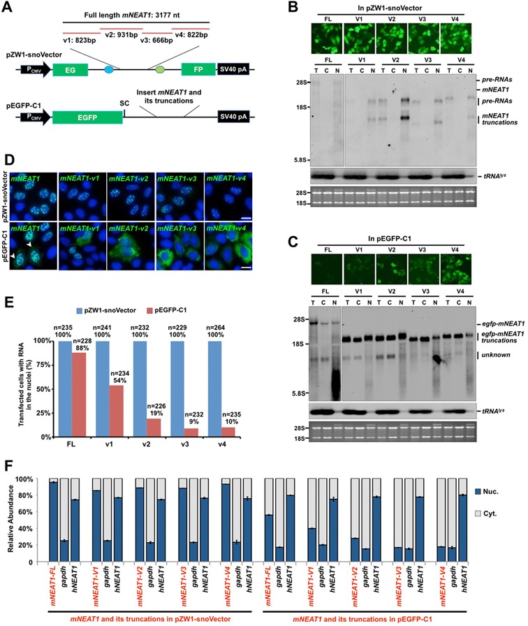
snoVectors express RNAs in the nucleus. (A) A schematic drawing to show full-length (FL) mNEAT1 RNA and different fragments of it (9) inserted into either the sno-lncRNA region in pZW1-snoVector (top) or into the UTR region of pEGFP-C1, which was engineered with a stop codon immediately downstream of the EGFP ORF (Bottom). (B) mNEAT1 RNA and different fragments of it expressed from pZW1-snoVector are retained in the nucleus. Top, HeLa cells were transfected with indicated plasmids individually and fluorescence pictures were taken 36 h after transfection. Bottom, total RNAs and fractionated nuclear and cytoplasmic RNAs were collected from the same batch of transfected HeLa cells as shown above, and then resolved on an agarose gel. Transcripts of mNEAT1 RNA and different fragments of it were probed with a Dig-labeled antisense SNORD116-13; tRNAlys was used as marker for nuclear/cytoplasmic RNA isolation. Equal amounts of total, cytoplasmic and nuclear RNAs were loaded onto an agarose gel and rRNAs were used as the loading control. T, total RNAs; C, cytoplasmic RNAs; N, nuclear RNAs. (C) mNEAT1 RNA and different fragments of it expressed from pEGFP-C1 are at least partially exported to the cytoplasm. Top, HeLa cells were transfected individually with the indicated plasmids. Total RNAs and fractionated nuclear and cytoplasmic RNAs were collected 36 h after transfection, and then resolved on an agarose gel. Transcripts of mNEAT1 RNA and different fragments of it were probed with Dig-labeled egfp. See (B) for details. (D) RNAs expressed from pZW1-snoVector are absolutely retained in the nucleus, while those from pEGFP-C1 are not. Top, HeLa cells were transfected with each indicated plasmid in a pZW1-snoVector for 36 h. RNA in situ hybridizations were performed with Dig-labeled probes for mNEAT1 RNA and different fragments of it. Bottom, HeLa cells were transfected with each indicated plasmid in a pEGFP-C1 vector for 36 h. RNA in situ hybridizations were performed with Dig-labeled egfp. Representative images are shown for each transfection. White arrow heads represent the cytoplasmic signals of egfp-mNEAT1-FL in cytoplasm in transfected HeLa cells. All nuclei were counterstained with DAPI. Scale bars 10 μm. (E) Subcellular distribution of transfected RNAs from different expression vectors. Quantitative analysis of the data from experiments shown in (D) is presented. More than 200 transfected cells were recorded randomly by confocal microscopy following each different transfection, and the percentage of each distinct nuclear/cytoplasmic localization pattern of RNAs transcribed from pZW1-snoVector or pEGFP-C1 was recorded. Note that mNEAT1 RNA and different fragments of it expressed from pZW1-snoVector are retained in the nucleus, while those from pEGFP-C1 are not. (F) mNEAT1 RNA and different fragments of it expressed from pZW1-snoVector are predominately retained in the nucleus. Nuclear and cytoplasmic RNAs extracted from equal numbers of transfected HeLa cells under each indicated condition were assayed by RT-qPCR. The cytoplasmic distributed gapdh and the nuclear retained endogenous hNEAT1 were used as markers to indicate a qualified cytoplasmic and nuclear fractionation under each transfection condition. Error bars were calculated from three replicates. In (B), (C) and (D), assays were repeated and the same results were obtained. Also see supplemental information Figure S2.