
Figure 3.
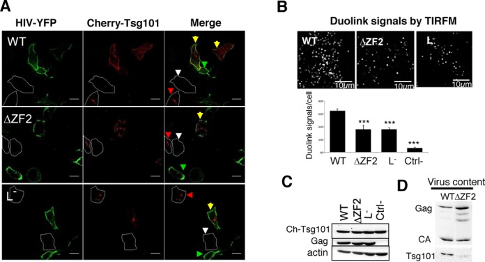
Recruitment of Tsg101 to HIV assembly sites and its incorporation into virions. (A) Representative confocal images of HeLa cells expressing mCherry-Tsg101 and untagged/tagged-YFP HIV (ratio 0.2:1:1). Yellow arrows show cells co-transfected with HIV and Tsg101, while green and red arrowheads show cells transfected only with HIV or Tsg101, respectively. White arrowheads indicate non-transfected cells. (B) Insitu Duolink experiments. Duolink immunolabeling generates fluorescent signals when Gag and Tsg101 proteins are in close proximity (≤40 nm). HeLa cells were cotransfected with cherry-Tsg101 and pNL4-3Δenv (WT, ΔZF2 or L−) for 20 h. A similar experiment was conducted without the anti-Tsg101 primary antibody as a negative control. Cells were imaged by TIRFM and the number of fluorescent Duolink spots per cell was measured using ImageJ. The histograms represent the average ± SD of the mean values of the Duolink signal per cell calculated from two independent experiments (n ≥ 27 cells) (more details in Text S7). ***P < 0.0001 using the Student's t-test. (C) Expression of Tsg101 and Gag proteins in cells used in Duolink assays was monitored by western blotting. (D) Incorporation of Tsg101 into virions. 293T cells were transfected with pNL4-3 (WT or ΔZF2) during 48 h. The production of virions was quantitated using p24 Elisa and normalized amounts of virions were loaded onto SDS-PAGE. Tsg101 and Gag proteins were detected by western blotting (Text S7).